[コンプリート!] e coli gram stain staphylococcus aureus 372463-How to gram stain e coli
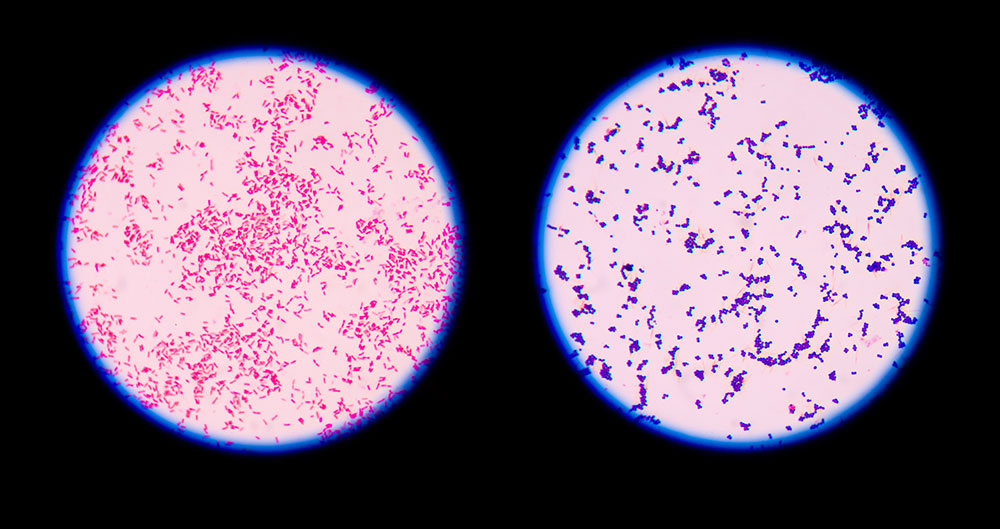
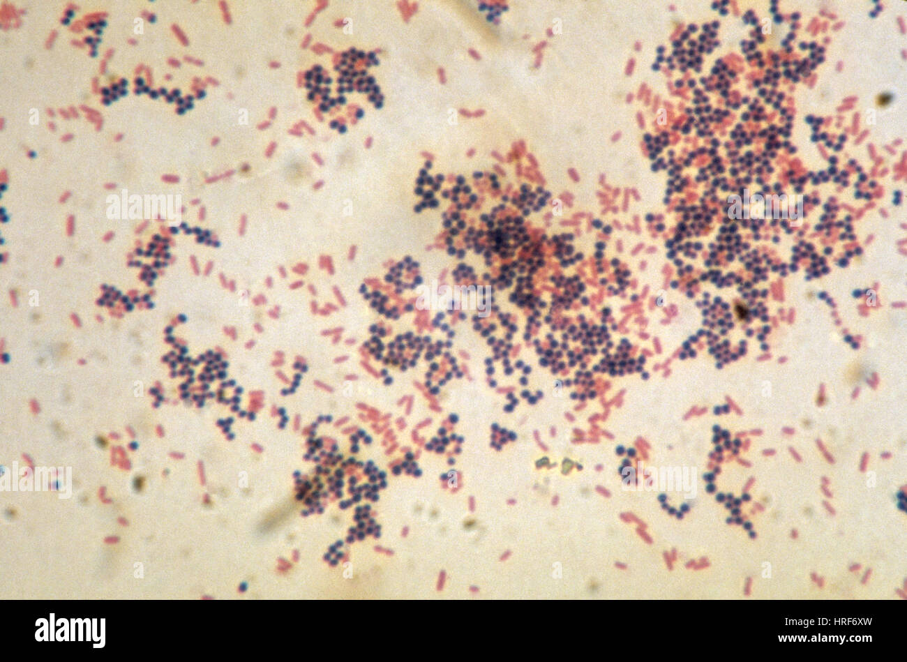
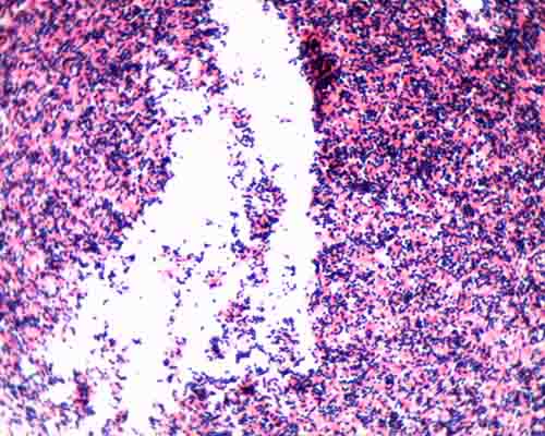
Microscopic Examination Unknown 52 was observed under oil immersion using the brightfield light microscope 1000x magnification after conducting Gram stain technique Escherichia coli (Gram negative rods) preprepared slide was the negative control while the Staphylococcus epidermidis (Gram positive cocci) preprepared slide provided theA Gram stain of mixed Staphylococcus aureus (S aureus ATCC , Grampositive cocci, in purple) and Escherichia coli (E coli ATCC , gramnegative bacilli, in red), the most common Gram stain reference bacteria Gram stain or Gram staining, also called Gram's method,Staphylococcus is a genus of Gram ve, round shape bacteria that are arranged in grapelike clusters The organisms of this genus are the commonest cause of suppurative lesions It was first isolated by Sir Alexander Ogston and he gave the name Staphylococcus for the typical arrangement of the cocci in grapelike clusters

Identifying Bacteria 1 Bms 503 Unh Elizabeth Valcourt Bms 504 05 28 September 17 So You Think You Can Identify Bacteria Introduction This Lab Was Studocu
How to gram stain e coli
How to gram stain e coli-Positive Gram Stain Negative E coli Serratia marcescens Proteus mirabilis Neisseria gonorrhea Alcaligenes Faecalis Proteus vulgaris Klebsiella pneumoniae Citrobacter freundii Pseudomonas fluorescens Enterobacter aerogenes Staphylococcus aureus Streptococcus pneumoniae Bacillus cereus Enterococcus faecalis Micrococcus luteus E coli SerratiaThe Gram stain is an example of a _____ stain, because the process uses two contrasting stains to separate bacteria into groups based on cell wall composition b E coli c Staphylococcus aureus Endospores What enabled the heat tolerance shown by the organism that survived the highest heat exposure?


Staphylococcus Aureus And Ecoli Under Microscope Microscopy Of Gram Positive Cocci And Gram Negative Bacilli Morphology And Microscopic Appearance Of Staphylococcus Aureus And E Coli S Aureus Gram Stain And Colony Morphology On Agar Clinical
False True/False Boiling water can bePDF Staphylococcus aureus and Escherichia coli are pathogens that can cause foodborne illnesses due to their ability to produce toxin These bacteria Find, read and cite all the researchStaphylococcus aureus and Escherichia coli were enumerated and isolated from readytoeat vegetables salad and meat luncheon on their selective media (Bairdparker and Macconkey agar, respectively) Twenty suspicious colonies of each (10 from each product) were randomly chosen and identified using conventional based on morphological and physiological characteristics
Diffusely adherent E coli (DAEC) Gram Stain E coli is described as a Gramnegative bacterium This is because they stain negative using the Gram stain The Gram stain is a differential technique that is commonly used for the purposes of classifying bacteria The staining technique distinguishes between two main types of bacteria (gramNewcomer Supply Gram Positive & Gram Negative Bacteria, Artificial Control Slides are for the positive histochemical staining of gram positive and gram negative bacteria in the same tissue section Escherichia coli and Staphylococcus aureus purchased from Remel Microbiology Products is used to produce the positive control tissueGramstain Grampositive cocci and Gramnegative bacilli Microscopic appearance Cocci in grapelike clusters (Saureus) and bacilli(Ecoli) Clinical significance of Saureus Frequently found as part of the normal skin flora on the skin and nasal passages;
PDF Staphylococcus aureus and Escherichia coli are pathogens that can cause foodborne illnesses due to their ability to produce toxin These bacteria Find, read and cite all the researchAmong them, Staphylococcus aureus (S aureus, SA) is one of the most prevalent Gram‐positive bacteria (Chao, et al, 15) S aureus strains are able to produce seven toxins responsible for multiple foodborne illnesses (Tenderis, et al, )Gram Staining Gram staining From Wikipedia, the free encyclopedia A Gram stain of mixed Staphylococcus aureus (Staphylococcus aureus ATCC , Grampositive cocci, in purple) andEscherichia coli (Escherichia coli ATCC , Gramnegative bacilli, in red), the most common Gram stain reference bacteria Gram staining (or Gram's method) is a method of differentiating bacterial species into


Microflora And Sanitary Indicative Bacteria Of The Soil Water Air The Methods Of Studying Online Presentation


E Coli Escherichia Staphylococcus Aureus Bacteria In Agar Plates Stock Photo Picture And Royalty Free Image Image
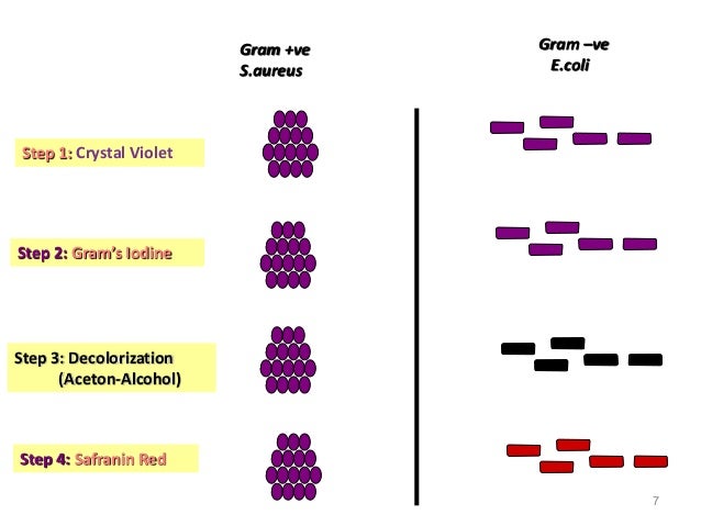
PDF Staphylococcus aureus and Escherichia coli are pathogens that can cause foodborne illnesses due to their ability to produce toxin These bacteria Find, read and cite all the researchGramstain Grampositive cocci and Gramnegative bacilli Microscopic appearance Cocci in grapelike clusters (Saureus) and bacilli(Ecoli) Clinical significance of Saureus Frequently found as part of the normal skin flora on the skin and nasal passages;Figure 234 In this specimen, the grampositive bacterium Staphylococcus aureus retains crystal violet dye even after the decolorizing agent is added Gramnegative Escherichia coli , the most common Gram stain qualitycontrol bacterium, is decolorized, and is only visible after the addition of the pink counterstain safranin


Antimicrobial Activity Of Lactic Acid Bacteria And The Spectrum Of Their Biopeptides Against Spoiling Germs In Foods


Www Mccc Edu Hilkerd Documents Bio1lab3 Exp 4 Pdf
Just like Ecoli, there are pathogenic strains of S Aureus that are responsible for skin infections, abscesses, and respiratory infections This bacteria is ideal for the gram staining technique since it is a grampositive bacteria, this means it has a thick peptidoglycan layer that will trap crystal violet and so will appear bluish/purpleCitric acid on E coli and S aureus were 006 g/mL, it was 003 g/mL for C albicans It was applied the TDtest with citric acid solution and discriminate by tolerance level of E coli than the other microorganisms at the end of the 48h incubation In a final test which was investigated the survival of E coli, S aureus and C albicansGram Staining Now that the slide has been heat fixed, you may now begin to stain the organism This is started by applying a few drops of crystal violet for 30 seconds, then rinsing with water for 5 seconds Next, cover the slide with Gram's Iodine and let sit for a full minute before rinsing with water for another 5 seconds


2


Solved 3 A Mixed Smear Of A 3 Day 72 Hour Culture Of S Chegg Com
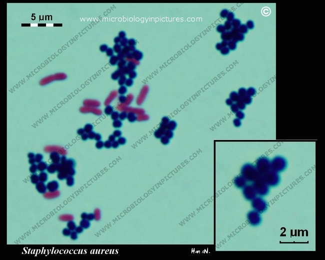
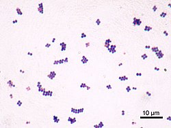
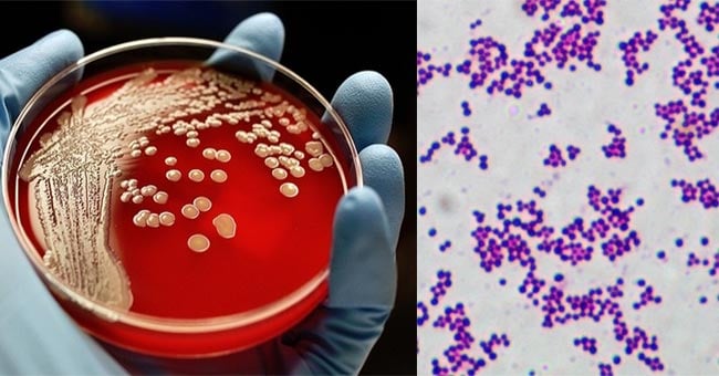
A direct smear for Gram staining may be performed as soon as the specimen is collected The Gram stain showing typical Grampositive cocci that occur singly and in pairs, tetrads, short chains, and irregular grapelike clusters can be suspected to be S aureus Image Source Microbiology in pictures and Sanmukh Joshi Culture Growth mediumStaphylococcus aureus is a Grampositive, roundshaped bacterium that is a member of the Firmicutes, and it is a usual member of the microbiota of the body, frequently found in the upper respiratory tract and on the skinIt is often positive for catalase and nitrate reduction and is a facultative anaerobe that can grow without the need for oxygen Although S aureus usually acts as a commensalPDF Staphylococcus aureus and Escherichia coli are pathogens that can cause foodborne illnesses due to their ability to produce toxin These bacteria Find, read and cite all the research


E Coli Vs S Aureus Gram Negative Vs Gram Positive Bacteria Youtube
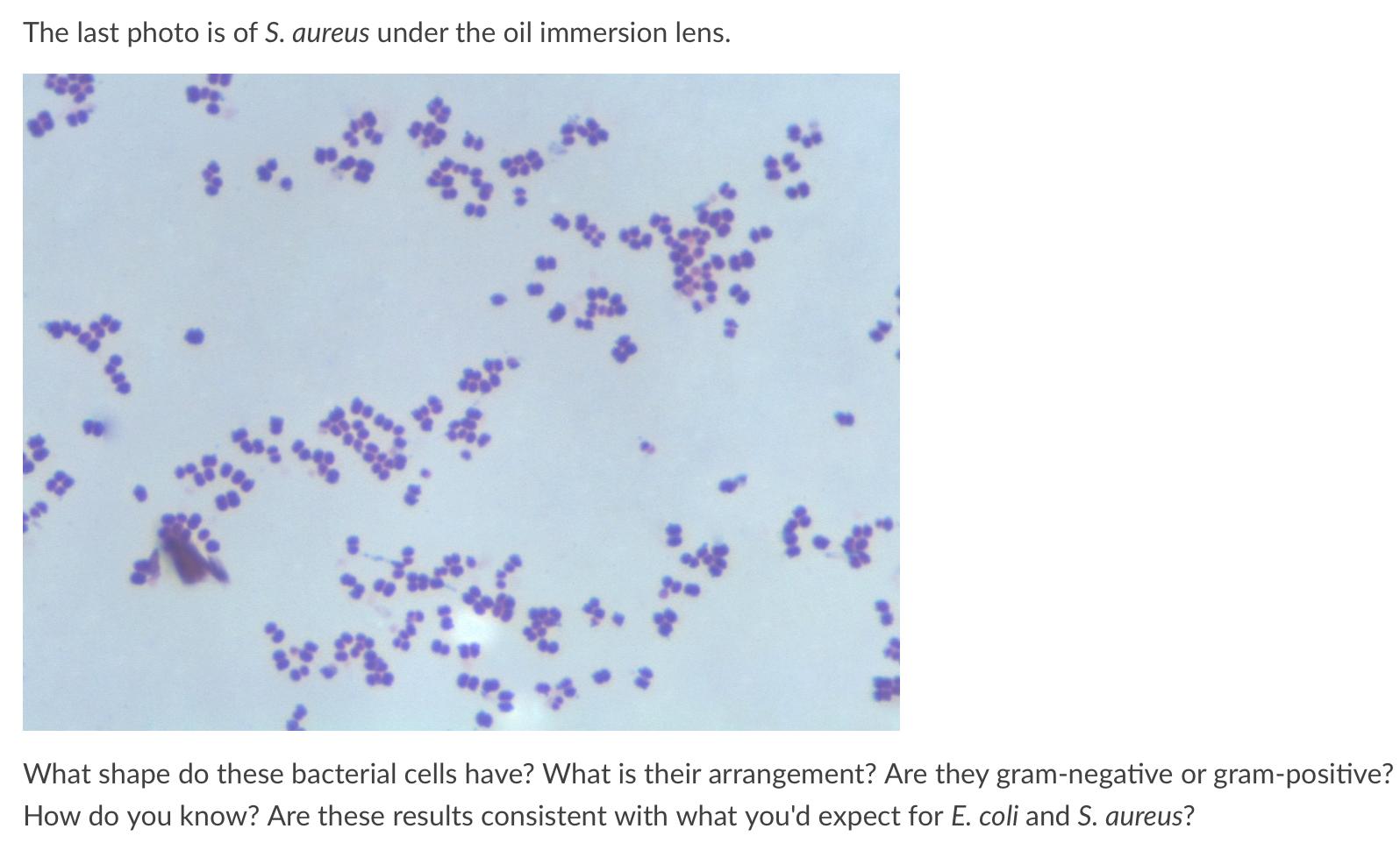


Solved The Last Photo Is Of S Aureus Under The Oil Immer Chegg Com
DIFFERENTIAL STAIN An example is Gram staining (or Gram's method) It is routinely used as an initial procedure in the identification of an unknown bacterial species Let's suppose we have a smear containing mixture of Staphylococcus aureus and Escherichia coli as in previous case We will use the same stains as before and besides we will needPositive Gram Stain Negative E coli Serratia marcescens Proteus mirabilis Neisseria gonorrhea Alcaligenes Faecalis Proteus vulgaris Klebsiella pneumoniae Citrobacter freundii Pseudomonas fluorescens Enterobacter aerogenes Staphylococcus aureus Streptococcus pneumoniae Bacillus cereus Enterococcus faecalis Micrococcus luteus E coli SerratiaOn staining, E coli appear as nonsporeforming, Gramnegative rodshaped bacterium Routine urine cultures should be plated using calibrated loops for the semiquantitative method Note The most commonly used criterion for defining significant bacteriuria is the presence of ⩾10 5 CFU per milliliter of urine


Staphylococcus Aureus And Ecoli Under Microscope Microscopy Of Gram Positive Cocci And Gram Negative Bacilli Morphology And Microscopic Appearance Of Staphylococcus Aureus And E Coli S Aureus Gram Stain And Colony Morphology On Agar Clinical


Gram Stain Tissue Trol Control Slides Mouse Lung Tissue Containing Staphylococcus Aureus And Escherichia Coli Sigma Aldrich
Gramstain Grampositive cocci and Gramnegative bacilli Microscopic appearance Cocci in grapelike clusters (Saureus) and bacilli(Ecoli) Clinical significance of Saureus Frequently found as part of the normal skin flora on the skin and nasal passages;It is estimated that % of the human population are longterm carriers of S aureusIt is estimated that % of the human population are longterm carriers of S aureus


E Coli Gram Stain Introduction Principle Procedure And Result Interpret


Pin On Mlt Stuff
DIFFERENTIAL STAIN An example is Gram staining (or Gram's method) It is routinely used asE coli is a gramnegative bacteriaS aureus is a grampositive coccus It is a coccus because its shape is round (from the Greek kokkos=grain)This is a mixed culture of Gram negative Escherichia coli (redorange) and Gram positive Staphylococcus aureus (bluepurple) stained using the Gram staining method Michael R Francisco/Flickr/CC BYSA


Microbiology Lab Flashcards Quizlet


Solved Microbe A 1 Which Of These Microbes Would Best Re Chegg Com
Bio 112 Abstract Escherichia coli, Bacillus subtilis and Staphylococcus epidermidis were analyzed for this lab activity to determine their Gram Stain After the multilayered Gram Stain procedure each bacteria were classified as Grampositive or Gramnegative depending on their cell walls staining color The results showed that E coli stained pink and classified as GramnegativeAbstract Staphylococcus aureus and Escherichia coli were enumerated and isolated from readytoeat vegetables salad and meat luncheon on their selective media (Bairdparker and Macconkey agar, respectively) Twenty suspicious colonies of each (10 from each product) were randomly chosen and identified using conventional based on morphological and physiological characteristicsStaphylococci (staphylococcus aureus)gram positive, spherical bacteris that have developed antibiotic resistant strains, 400x gram stain stock pictures, royaltyfree photos & images microscopic view of bacterial pneumonia bacterial pneumonia is a type of pneumonia caused by bacterial infection gram stain stock illustrations


9 Gram Staining Best Practices Microbiologics Blog


Q Tbn And9gcqrzvobmfeiajtjsl54jck6oeivvjvclzt9utdfbxfiq3swwfrw Usqp Cau
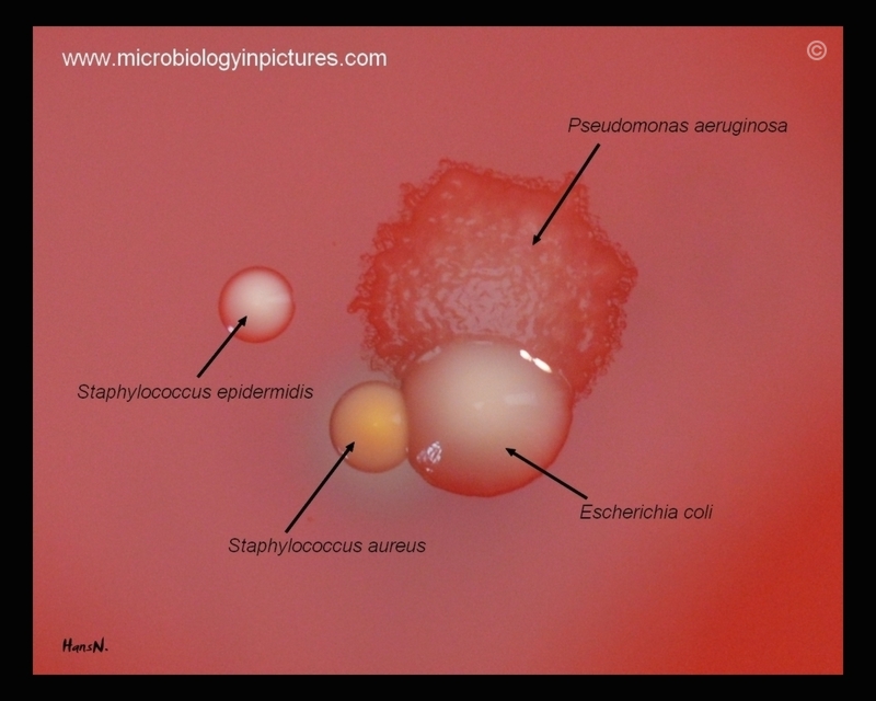
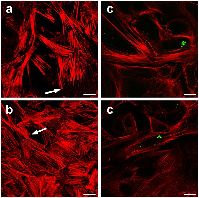
Temperature • Psychrophiles (150 C) – Pseudomonas fluorescens • Mesophiles (400 C) – Escherichia coli, Salmonella enterica, Staphylococcus aureus • Thermophiles ( C) Bacillus stearothermophilus • Extremely thermophiles (as high as 2500 C) 16 17Staphylococcus aureus and Escherichia coli are among the most prevalent species of grampositive and gramnegative bacteria, respectively, that induce clinical mastitis The innate immune system comprises the immediate host defense mechanisms to protect against infection and contributes to the initial detection of and proinflammatory responseStaphylococcus aureus Staphylococcus aureus on Columbia agar with 5% defibrinated sheep blood (BioRad™) Individual colonies on agar are round, convex, and 14 mm in diameter with a sharp borderOn blood agar plates, colonies of Staphylococcus aureus are frequently surrounded by zones of clear betahemolysisThe golden appearance of colonies of some strains is the etymological root of the


Fesem Images Of Gram Negative E Coli And Gram Positive S Aureus On Download Scientific Diagram


Prokaryotic Cell Anatomy
Jan 29, 14 Staphylococcus sp (simple stain, methylene blue)It is estimated that % of the human population are longterm carriers of S aureusChlorhexidine prevents the transformation of Bacillus megaterium cells to protoplasts by lysozyme, of E coli cells to spheroplasts by penicillin and causes lysis of "protoplasts" and spheroplasts of E coli stabilised in hypertonic sucrose solution Transformation of Staphylococcus aureus cells to the Gram‐negative condition occurred in contact with 10 to 800 μg/ml of chlorhexidine and


Bacteria Exercise Flashcards Quizlet
/gram-positive-staphylococcus-aureus-bacteria-541802136-57979cca5f9b58461f26eccc.jpg)


Gram Stain Procedure In Microbiology
E coli bacteria are gramnegative, so they stain pink in a gram test E coli bacteria were first identified by bacteriologist Dr Escherich in 15 Since then, more than 700 different serotypes have been identified Some of these strains are pathogenic, but most are not A strain known as uropathogenic E coli, or UPEC, is known to causeThe Gram stain is an example of a _____ stain, because the process uses two contrasting stains to separate bacteria into groups based on cell wall composition b E coli c Staphylococcus aureus Endospores What enabled the heat tolerance shown by the organism that survived the highest heat exposure?Staphylococcus aureus and Escherichia coli were enumerated and isolated from readytoeat vegetables salad and meat luncheon on their selective media (Bairdparker and Macconkey agar, respectively) Twenty suspicious colonies of each (10 from each product) were randomly chosen and identified using conventional based on morphological and physiological characteristics


Science Source Stock Photos Video Escherichia Coli And Staph Aureus


Gram Stain Staphylococcus Aureus And Escherichia Coli Gram Staining Technique Micrograph Of S Aureus And E Coli
Gramstain, Staphylococcus aureus and Escherichia coli Gram staining technique, micrograph of Saureus and Ecoli Mixture of Grampositive cocci ( Staphylococcus aureus) and Gramnegative rods ( Escherichia coli ) Gram staining technique, magnification ×1000The effect of NaCl concentration on the growth of gram negative Escherichia coli and gram positive Staphylococcus aureus cells cultivated at 37°C was studied in an effort to understand theJust like Ecoli, there are pathogenic strains of S Aureus that are responsible for skin infections, abscesses, and respiratory infections This bacteria is ideal for the gram staining technique since it is a grampositive bacteria, this means it has a thick peptidoglycan layer that will trap crystal violet and so will appear bluish/purple


Staphylococcus Aureus And Ecoli Under Microscope Microscopy Of Gram Positive Cocci And Gram Negative Bacilli Morphology And Microscopic Appearance Of Staphylococcus Aureus And E Coli S Aureus Gram Stain And Colony Morphology On Agar Clinical
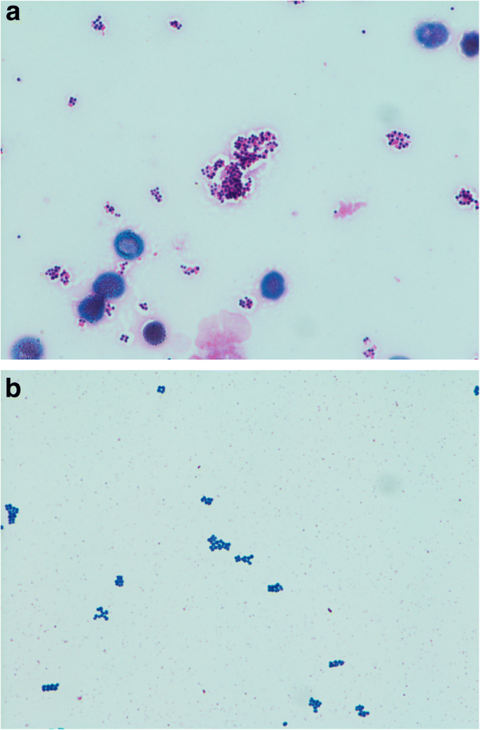


Validation Of Blood Culture Gram Staining For The Detection Of Staphylococcus Aureus By The Oozing Sign Surrounding Clustered Gram Positive Cocci A Prospective Observational Study Bmc Infectious Diseases Full Text
Bio 112 Abstract Escherichia coli, Bacillus subtilis and Staphylococcus epidermidis were analyzed for this lab activity to determine their Gram Stain After the multilayered Gram Stain procedure each bacteria were classified as Grampositive or Gramnegative depending on their cell walls staining color The results showed that E coli stained pink and classified as Gramnegative Both B subtilis and S epidermidis stained purple and were classified as Grampositive It was determined that EcStaphylococcus aureus and Escherichia coli are among the most prevalent species of grampositive and gramnegative bacteria, respectively, that induce clinical mastitis The innate immune system comprises the immediate host defense mechanisms to protect against infection and contributes to the initial detection of and proinflammatory response to infectious pathogensFalse True/False Boiling water can be


Identifying Bacteria 1 Bms 503 Unh Elizabeth Valcourt Bms 504 05 28 September 17 So You Think You Can Identify Bacteria Introduction This Lab Was Studocu
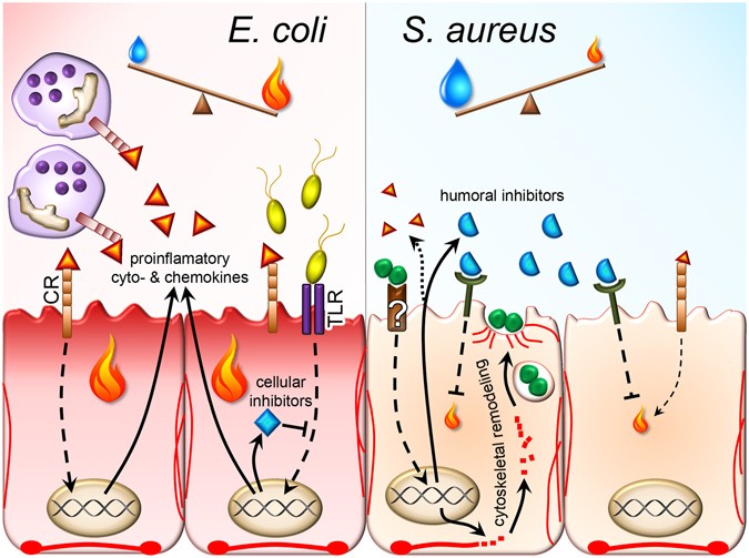


Differentiating Staphylococcus Aureus From Escherichia Coli Mastitis S Aureus Triggers Unbalanced Immune Dampening And Host Cell Invasion Immediately After Udder Infection Scientific Reports
Altogether, both pyridoxine derivatives successfully inhibited the growth of grampositive bacteria (Staphylococcus aureus, Staphylococcus epidermidis, Bacillus subtilis) and Escherichia coli considerably, while demonstrated nearly no effect against Klebsiella pneumoniae and Pseudomonas aeruginosaB) A Gram stain of grampositive cocci Staphylococcus aureus (violet or purple) and the gramnegative bacilli Escherichia coli (pink), which are the most commonly used Gram stain reference bacteria Adapted from Wikipedia commons)The experiment verified that E coli is Gramnegative while S aureus is the genus of Grampositive bacteria In the conducted Gram stain analysis, the main coloring agent actively bound with the murein layer


Experiment 4a Lab04 Virtual Edge Molb 21 College Of Agriculture And Natural Sciences


Product Catalog
B) A Gram stain of grampositive cocci Staphylococcus aureus (violet or purple) and the gramnegative bacilli Escherichia coli (pink), which are the most commonly used Gram stain reference bacteria Adapted from Wikipedia commons)A direct smear for Gram staining may be performed as soon as the specimen is collected The Gram stain showing typical Grampositive cocci that occur singly and in pairs, tetrads, short chains, and irregular grapelike clusters can be suspected to be S aureus Image Source Microbiology in pictures and Sanmukh Joshi Culture Growth mediumThe main purpose of this study was to explore the antibacterial activity and mechanism of lacidophilin from Lactobacillus pentosus against Staphylococcus aureus and Escherichia coli The effects of temperature, enzyme, metal ions, and pH on the antibacterial activity of L pentosus were evaluated The result showed that lacidophilin had good thermal stability and could be decomposed by trypsin


Gram Stain


Gram Stain Demonstration Slide 400x 1 Marc Perkins Photography
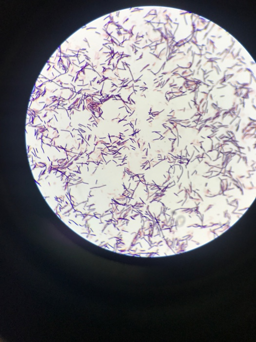


Solved Where Is The Gram Staining Mix Staphylococcus Aure Chegg Com


Gram Negative E Coli And Gram Positive S Aureus Bacterial Growth Download Scientific Diagram


Modification Of Membrane Properties And Fatty Acids Biosynthesis Related Genes In Escherichia Coli And Staphylococcus Aureus Implications For The Antibacterial Mechanism Of Naringenin Sciencedirect


Agar Plate Images Left Staphylococcus Aureus Gram Positive And Download Scientific Diagram


Gram Stain Staphylococcus Aureus E Coli Combined In Same Organ Control Histology Slides


General Microbiology Staining Bacteria Cells Staining Bacteria Cells


Gram Stain Lab Report Record The Results Use The F Chegg Com
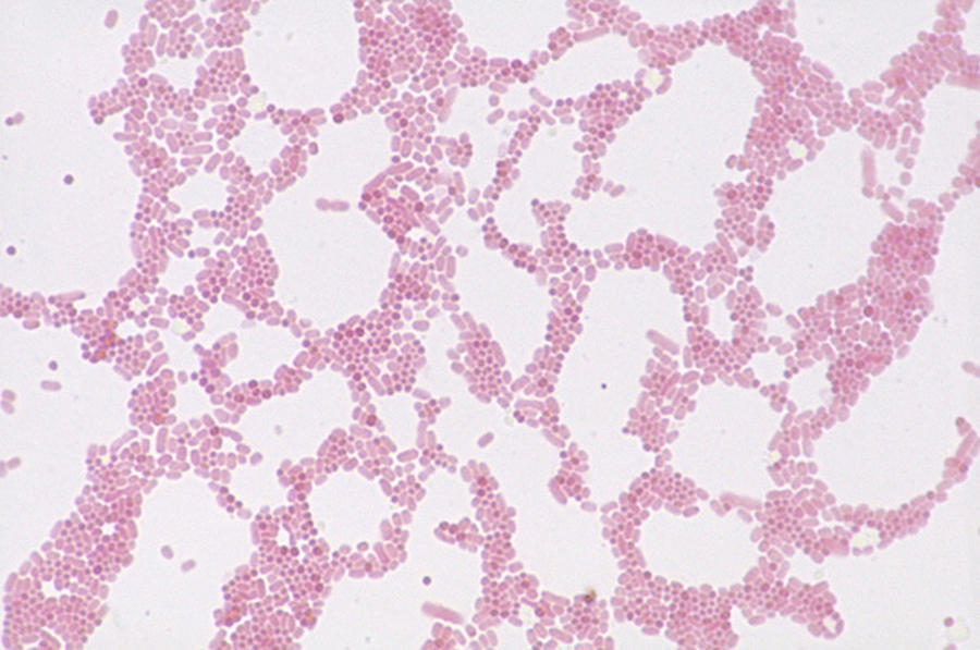


Escherichia Coli And Staph Aureus Photograph By Michael Abbey
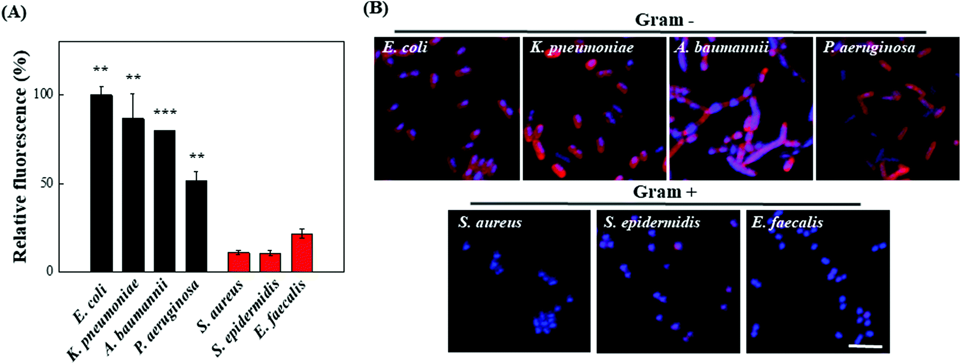


Ultra Fast And Universal Detection Of Gram Negative Bacteria In Complex Samples Based On Colistin Derivatives Biomaterials Science Rsc Publishing


Gram Staining Of Isolated Clinical Strains A S Aureus B S Download Scientific Diagram


Gram Stained Micrograph High Resolution Stock Photography And Images Alamy


Gram Stain Wikipedia


Q Tbn And9gcqkye60ou Johpr02n Mbv1fferrjpdh Lnct7ymdf5qhyia1ld Usqp Cau


Escherichia Coli Colony Morphology And Microscopic Appearance Basic Characteristic And Tests For Identification Of E Coli Bacteria Images Of Escherichia Coli Antibiotic Treatment Of E Coli Infections


Gram Stain Ppt Download


Solved 10 M If This Is A Gram Stain Of Staphylococcus Aur Chegg Com


Gram Stain Microbiology Images Photographs


Www Mccc Edu Hilkerd Documents Bio1lab3 Exp 4 Pdf


Staphylococcus Aureus And Ecoli Under Microscope Microscopy Of Gram Positive Cocci And Gram Negative Bacilli Morphology And Microscopic Appearance Of Staphylococcus Aureus And E Coli S Aureus Gram Stain And Colony Morphology On Agar Clinical


E Coli Escherichia Image Photo Free Trial Bigstock


Staphylococcus Aureus Wikipedia


Journal Of Microbiology And Antimicrobials Bactericidal Activities Of The Leaf Extract Of Spondias Mombin L Plant Used For Culinary And Therapeutically Purposes In Ivory Coast


Www Mccc Edu Hilkerd Documents Bio1lab3 Exp 4 Pdf


Staphylococcus Aureus Disease Properties Pathogenesis And Laboratory Diagnosis Learn Microbiology Online


Gram Staining Miceobiology Student


Q Tbn And9gctgrmjg8acjrbxbniknzy1qewngtoobg8cixbwsv Ok9ny02ti0 Usqp Cau


Gram Stain Staphylococcus Aureus E Coli Combined In Same Organ Control Histology Slides


In Vitro Study Of The Activity Of Some Medicinal Plant Leaf Extracts On Urinary Tract Infection Causing Bacterial Pathogens Isolated From Indigenous People Of Bolangir District Odisha India Biorxiv


Gram Stain


Gram S Staining Of S Aureus 100x Grapes Like Black Arrow Gram Download Scientific Diagram


E Coli P Aeruginosa S Aureus And S Epidermidis Bacteria Colonies On Blood Agar


Biochemical Test Of Staphylococcus Aureus Microbe Notes


Evaluation Of The Antimicrobial Activity Of Chitosan And Its Quaternized Derivative On E Coli And S Aureus Growth


Q Tbn And9gctgrmjg8acjrbxbniknzy1qewngtoobg8cixbwsv Ok9ny02ti0 Usqp Cau


Differentiating Staphylococcus Aureus From Escherichia Coli Mastitis S Aureus Triggers Unbalanced Immune Dampening And Host Cell Invasion Immediately After Udder Infection Scientific Reports


Bacteria Diversity Of Structure Of Bacteria Britannica


Kimicontrol Educational Covid 19 H1n1 H5n1 Legionella Ebola Pneumophilastaphylococcus Aureus Bacillus Anthracis Escherichia Coli Salmonella Listeria Monocytogenes Clostridium Botulinum Bacillus Cereus Yersinia Entercolitica Pseudomonas Aeruginosa


Bacterial Microscopy Streaked Images


About Science Prof Online Ppt Video Online Download


Solved 9 Identify The Gram Positive Or Gram Negative Sta Chegg Com


E Coli Escherichia Staphylococcus Aureus Bacteria In Agar Plates Stock Photo Picture And Royalty Free Image Image


File Escherichia Coli And Staph Aureus Jpg Wikimedia Commons
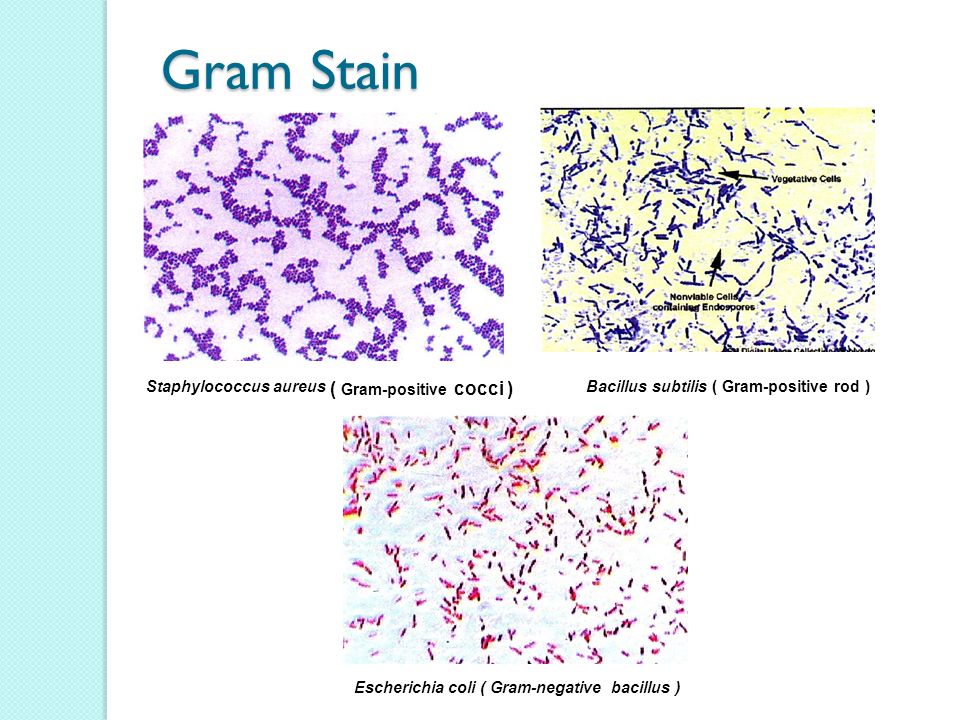


Observation Of Bacteria Using Staining Procedures Ppt Video Online Download


Two Petri Plates With Growing Bacterial Cultures In Medical Laboratory Stock Image Image Of Agar Infection


2 3 The Peptidoglycan Cell Wall Biology Libretexts
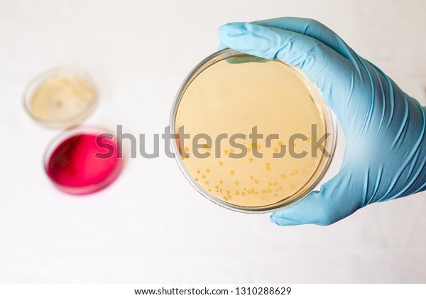


Hand Holds Petri Dish Ecoli Escherichia Stock Photo Edit Now


52 Microbiology Unknown Project Cscc Bio 2215 Ideas Microbiology Medical Laboratory Medical Laboratory Science


Utis Definition Infectious Diseases Of The Urinary Tract Ppt Video Online Download


Microlab Tutor Identification Of Unknown Bacteria Flashcards Quizlet


Gram Staining Procedure 42 Rights Managed Stock Photo Corbis Microbiology Medical Laboratory Educational Materials
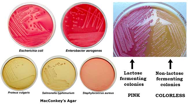


Macconkey Agar Composition Principle Uses Preparation And Colony Morphology
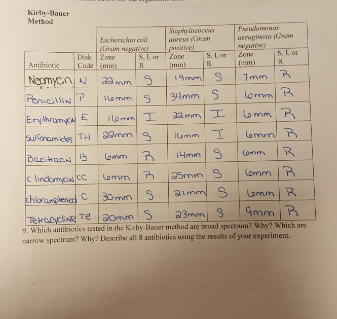


Solved Kirby Bauer Method Escherichia Coli Gram Negative Chegg Com


Bacterial Microscopy Streaked Images


Bacteria Overview Amboss


Effect Of Hydrogel On Gram Negative And Positive Bacteria By Download Scientific Diagram


Staphylococcus Aureus And Ecoli Under Microscope Microscopy Of Gram Positive Cocci And Gram Negative Bacilli Morphology And Microscopic Appearance Of Staphylococcus Aureus And E Coli S Aureus Gram Stain And Colony Morphology On Agar Clinical


Morphology Of Bacterial Cells


Solved 2 10mm Figure 1 Gram Stain Bacteria Are Escherich Chegg Com


Ex 8 Gram Stain Scientist Cindy


Solved Pea E Coli S Aureus Question 1 You Accidently M Chegg Com


Bacterial Microscopy Streaked Images


Gram Stained S Aureus In The Absence Of Agnps A Gram Stained S Download Scientific Diagram


In Gram S Staining A Gramnegative E Coli B Gram Negative Download Scientific Diagram
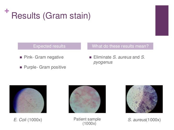


Case Study 1 Peanut Butter Is Tainted


Identification Of S Aureus And E Coli From Clinical Specimens Download Table


Bacterial Microscopy Streaked Images


Mics Of Peptide Analogs Against Gram Negative E Coli Ml35p And Download Table
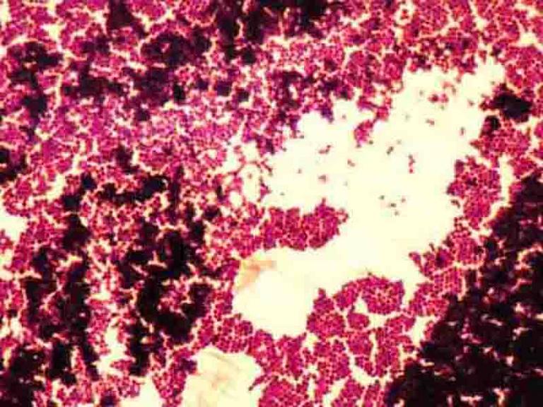


Gram Stain Microbiology Images Photographs


Evaluation Of The Antimicrobial Activity Of Chitosan And Its Quaternized Derivative On E Coli And S Aureus Growth Sciencedirect


Gram S Staining Of S Aureus 100x Grapes Like Black Arrow Gram Download Scientific Diagram
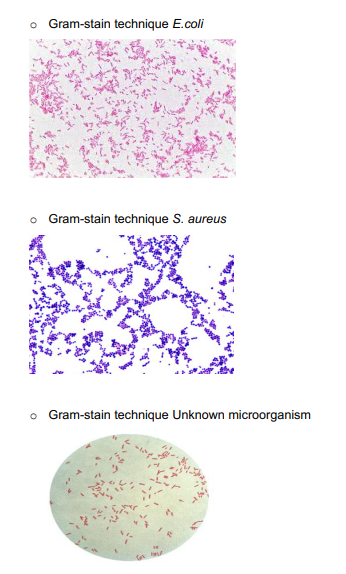


Solved For Each Image Describe The Bacteria S Color The Chegg Com


Gram Stain
2.jpg)


Biol 2


Gram Stain Demonstration Slide 400x 2 Marc Perkins Photography


コメント
コメントを投稿